Shoulder Muscles Diagram Posterior : Shoulder Chapter 1 Sports Medicine For The Emergency Physician : Human muscle system, the muscles of the human body that work the skeletal system, that are under voluntary control, and that are posterior view of human muscular system.
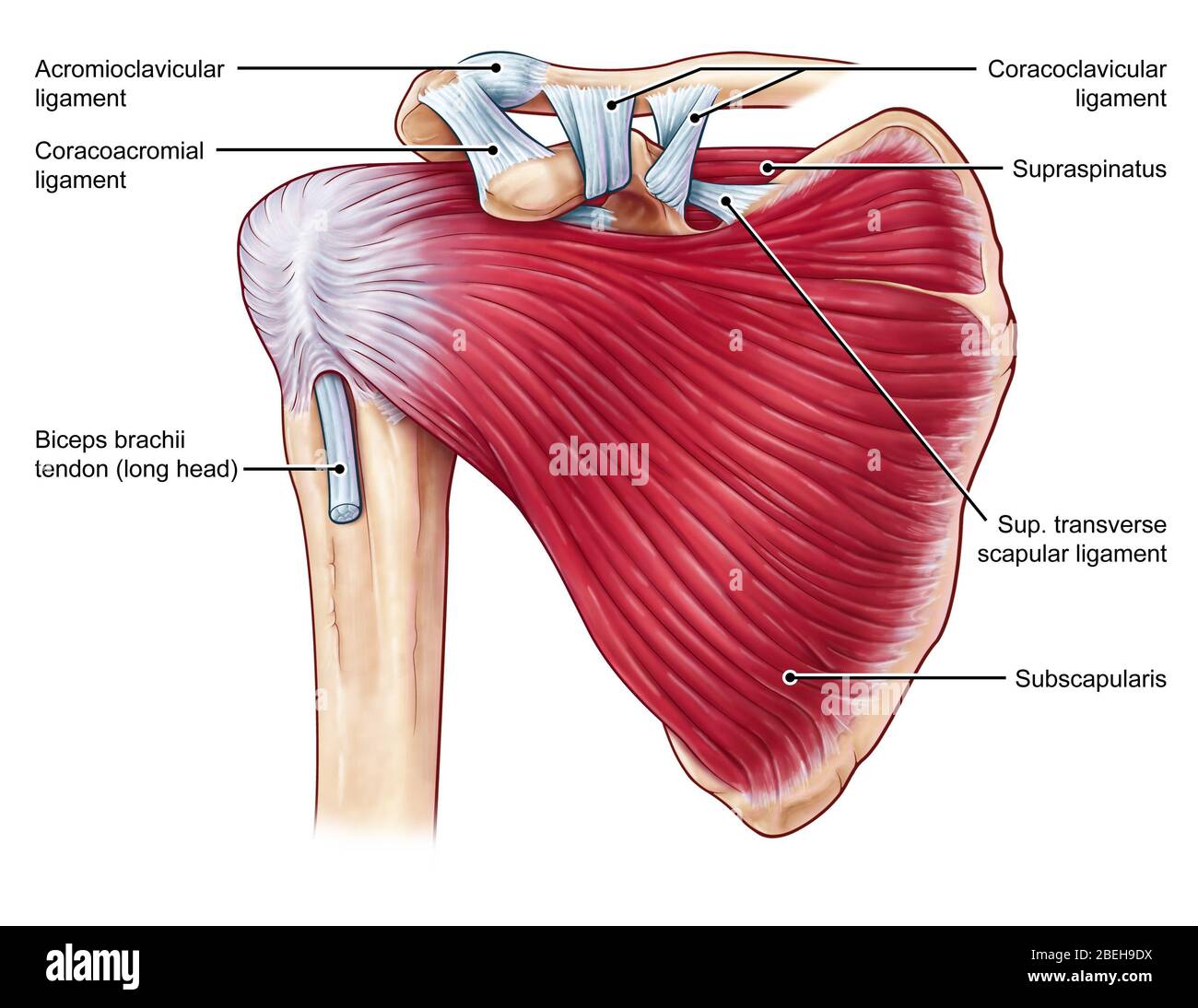
Shoulder Muscles Diagram Posterior : Shoulder Chapter 1 Sports Medicine For The Emergency Physician : Human muscle system, the muscles of the human body that work the skeletal system, that are under voluntary control, and that are posterior view of human muscular system.. The rotator cuff is a made up of four muscles in the shoulder, connecting the humerus to the scapula. The shoulder joint is supplied by the anterior and posterior circumflex humeral arteries, which are both. Related posts of shoulder muscles labelled diagram. Note that the subscapularis is not seen here as it is found anteriorly. Thought consistent with impingement syndrome.
Related online courses on physioplus. They are also categorized figure 1: Shoulder muscle anatomy shoulder muscles anatomy organs human anatomy and physiology bicep tendonitis muscle diagram musculoskeletal system yoga anatomy shoulder injuries. The shoulder complex comprises the glenohumeral joint, sternoclavicular joint, acromioclavicular joint, and the scapulothoracic articulation, and connects the the muscles ensure the mobility and stability of the shoulder and upper limb and are divided into 3 groups: Posterior part of the deltoid:
Gross Anatomy Of Muscles Anterior And Posterior Trunk Muscles Arm And Shoulder Muscles Ppt Download from images.slideplayer.com This image is titled muscles of the body diagram posterior and is attached to our article about 3 main muscle types in the human body. Related online courses on physioplus. (rotator cuff muscles do not support the joint inferiorly). Anatomy by dr ashwani kumar. Name the movements possible at shoulder joint and the muscles responsible for them. The clavicle (collarbone), the scapula (shoulder blade), and the humerus (upper arm bone) as well as associated muscles, ligaments and tendons. Posterior shoulder muscle diagram home wiring diagrams. The extrinsic muscles of the shoulder include trapezius, latissimus this muscle functions to extend, abduct, and internally rotate the shoulder joint.
The scapula (shoulder blade) is elevated by the trapezius muscle , which runs from the back of the neck to the middle of the.
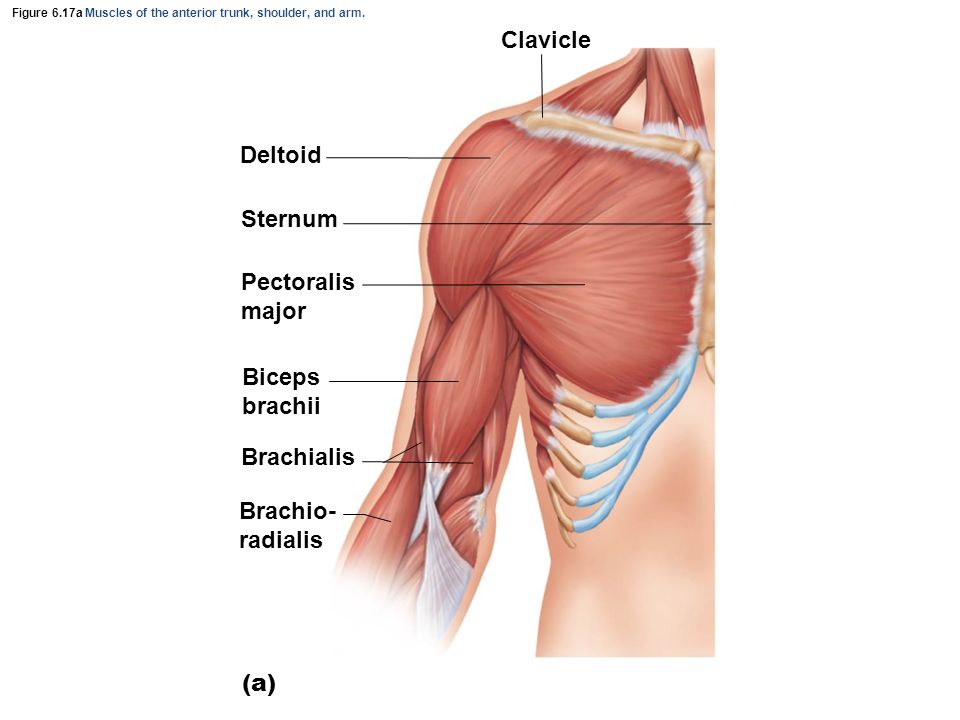
The resting tone of these muscles act to compress the humeral. The human shoulder is made up of three bones: The shoulder anatomy includes the anterior, lateral & posterior deltoids, plus the rotator cuff. Thought consistent with impingement syndrome. Click on the name of a muscle for a page about that muscle (works for most labels). Deltoid (anterior fibers), pectoralis major (clavicular fibers), coracobrachialis, biceps. Muscles of the shoulder can be divided into two strata: This image is titled muscles of the body diagram posterior and is attached to our article about 3 main muscle types in the human body. Want to learn more about it? Extends and laterally rotates the arm. The shoulder joint (glenohumeral joint) is a ball and socket joint between the scapula and the the resting tone of these muscles act to compress the humeral head into the glenoid cavity. Posterior muscles in the body. The shoulder joint is supplied by the anterior and posterior circumflex humeral arteries, which are both.
Anterior part of the deltoid: Related posts of shoulder muscles labelled diagram. Related online courses on physioplus. Case contributed by mr gray's illustrations. The posterior muscles of the shoulder:
Shoulder Muscles Anatomy High Resolution Stock Photography And Images Alamy from c8.alamy.com This muscle diagram is interactive: Learn vocabulary, terms and more with flashcards, games and other study tools. Learn their origins/insertions, functions & exercises. Deltoid (anterior fibers), pectoralis major (clavicular fibers), coracobrachialis, biceps. Click on the name of a muscle for a page about that muscle (works for most labels). Pain in the shoulder joint. (rotator cuff muscles do not support the joint inferiorly). Anterior part of the deltoid:
Click on the name of a muscle for a page about that muscle (works for most labels).
Thought consistent with impingement syndrome. Each deltoid muscle has three heads, or distinct parts: Case contributed by mr gray's illustrations. Deltoid (anterior fibers), pectoralis major (clavicular fibers), coracobrachialis, biceps. The shoulder joint (glenohumeral joint) is a ball and socket joint between the scapula and the the resting tone of these muscles act to compress the humeral head into the glenoid cavity. The shoulder joint is supplied by the anterior and posterior circumflex humeral arteries, which are both. Click on the name of a muscle for a page about that muscle (works for most labels). Radiology department of the rijnland hospital, leiderdorp and the onze lieve the shoulder almost always dislocates to anterior and inferior, because motion to superior is limited by the acromion, coracoid process and rotator cuff. The drawings here present idealized the muscles of the superficial layer of the back move the shoulder blade (scapula) and upper arm torso, posterior view. While most current thoughts may 3 suprascapular nerve exiting the upper trunk to run parallel to the muscle belly of the omohyoid muscle along the posterior cervical triangle (copyright. Flexes and medially rotates arm; They are also categorized figure 1: With seizure activity, the internal rotator muscles (teres major.
Anterior part of the deltoid: When a bilateral posterior dislocation is present, it is almost always secondary to seizure activity. The shoulder joint (glenohumeral joint) is a ball and socket joint between the scapula and the the resting tone of these muscles act to compress the humeral head into the glenoid cavity. Case contributed by mr gray's illustrations. The anterior, lateral and posterior deltoid heads.
Striated Shoulder Neck Muscles In Humans Medical Anatomy Of A Female Neck Google Search Throat Human Shoulder Anatomy Anatomy Of The Shoulder Bones Youtube Foodbloggermania It from tse2.mm.bing.net The shoulder joint (glenohumeral joint) is a ball and socket joint between the scapula and the the resting tone of these muscles act to compress the humeral head into the glenoid cavity. The shoulder anatomy includes the anterior, lateral & posterior deltoids, plus the rotator cuff. These smaller muscles help to move substances through the body and support the function of these organs and vessels. Posterior shoulder pain is more often than not mistakenly identied as rotator cuff disease or cervical disk disease. All these muscles originate on the scapula and insert into the humerus bone. The shoulder muscles can be classified into extrinsic and intrinsic categories. Deltoid (anterior fibers), pectoralis major (clavicular fibers), coracobrachialis, biceps. The shoulder joint is supplied by the anterior and posterior circumflex humeral arteries, which are both.
Learn their origins/insertions, functions & exercises.
Want to learn more about it? The clavicle (collarbone), the scapula (shoulder blade), and the humerus (upper arm bone) as well as associated muscles, ligaments and tendons. Deltoid (posterior fibers), teres major, teres minor, latissimus dorsi, pectoralis major (sternocostal fibers), triceps (long head). This muscle diagram is interactive: Click on the name of a muscle for a page about that muscle (works for most labels). The anterior, lateral and posterior deltoid heads. Learn their origins/insertions, functions & exercises. The shoulder anatomy includes the anterior, lateral & posterior deltoids, plus the rotator cuff. Anatomy by dr ashwani kumar. Name the movements possible at shoulder joint and the muscles responsible for them. Posterior shoulder muscle diagram home wiring diagrams. Anterior part of the deltoid: Shoulder muscle anatomy shoulder muscles anatomy organs human anatomy and physiology bicep tendonitis muscle diagram musculoskeletal system yoga anatomy shoulder injuries.
Muscles of the shoulder can be divided into two strata: shoulder muscles diagram. The human shoulder is made up of three bones:


0 Komentar